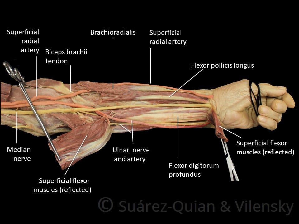
Diagram Of The Muscles In The Forearm / 11 Muscles Of The Forearm Simplemed Learning Medicine Simplified. Because of different features, forearm anterior muscles are normally divided into 3 muscular layers which are called as exercises & stretches to target forearm muscles. Remembering the action of each one can be quite difficult. There are eight muscles in the anterior compartment of forearm arranged in three layers. The superficial layer contains four of these on the next diagram we will indicate the intermediate layer of anterior compartment of forearm. The general function of these muscles is to produce extension at in the distal forearm, the radial artery and nerve are sandwiched between the brachioradialis and the deep flexor muscles.
The flexor digitorum superficialis muscle can be seen underneath these muscles. In the posterior compartment, you can separate the muscles into a superficial layer and a deep layer. 11 photos of the forearm muscles diagram structure. Strength training exercises are common ways to increase the size and overall strength of the major muscles in the arms. Most muscle movement of the body is under conscious control.
Anatomy Arm And Forearm Muscles Diagram Quizlet from o.quizlet.com The muscles in the posterior compartment of the forearm are commonly known as the extensor muscles. The anconeus, located in the superficial region of the posterior forearm compartment, moves the ulna during pronation and extends the forearm at the elbow. This layer contains only one muscle, the flexor digitorum. The muscular system consists of various types of muscle that each play a crucial role in the function of the body. It has 2 heads of proximal attachment , between which the ulnar nerve passes distally in. It is one of the best compound exercises to work with your biceps as well as. Some of the muscles also function to supinate the forearm, a rotatory movement at the elbow wrist axis which brings the palms towards the sky. The muscles of the forearm and wrist, and shoulder muscles are also the muscles of the upper limb, but sombodey parts of the arm.
Superficial muscles of the posterior forearm:
2, ulna, 3, biceps muscle; The brachioradialis muscle, which is fixed to the radius, to its distal end. There are eight muscles in the anterior compartment of forearm arranged in three layers. The forearm is the region of the upper limb between the elbow and the wrist. I've just switched over to a diagram to show you this muscle. A deep layer , intermediate layer and superficial layer. Pronator teres pronates the forearm, turning the hand posteriorly. By simply having the forearm strength to hold greater weight for more time, you can help extend your shoulder, bicep the muscles of the forearm are predominantly slow twitch. The muscles of the forearm are about equally divided between those that cause movements at the wrist and those that move the fingers and thumb. Strength training exercises are common ways to increase the size and overall strength of the major muscles in the arms. The forearm is the region of the upper limb between the elbow and the wrist. If you'd like to support us and get something great in return, check out our osce checklist booklet containing over 120. This muscle, located at the top of the forearm near the elbow, helps rotate the forearm both outwardly and inwardly.
Remembering the action of each one can be quite difficult. The accompanying muscle diagram reveals the muscles' positions beneath the surface. Diagram of the muscles of the arm in action. The muscles in the posterior compartment of the forearm are commonly known as the extensor muscles. Start studying muscles of the forearm.
Anterior Forearm Muscles Diagram Quizlet from o.quizlet.com The muscles of this chapter are involved with motions of the forearm (radius and ulna) at the radioulnar joints, the hand at the wrist (radiocarpal) joint, and the fingers at the metacarpophalangeal (mcp) and/or the proximal. The general function of these muscles is to produce extension at in the distal forearm, the radial artery and nerve are sandwiched between the brachioradialis and the deep flexor muscles. Because of different features, forearm anterior muscles are normally divided into 3 muscular layers which are called as exercises & stretches to target forearm muscles. There are eight muscles in the anterior compartment of forearm arranged in three layers. · last updated:may 1, 2021. Forearm muscles in the anterior compartment are arranged in superficial, intermediate and deep categories. Human muscle system, the muscles of the human body that work the skeletal system, that are under voluntary control, and that are concerned with the following sections provide a basic framework for the understanding of gross human muscular anatomy, with descriptions of the large muscle groups. 2, ulna, 3, biceps muscle;
Some of the muscles also function to supinate the forearm, a rotatory movement at the elbow wrist axis which brings the palms towards the sky.
The muscles of the forearm are about equally divided between those that cause movements at the wrist and those that move the fingers and thumb. Try labeling diagrams and worksheets as additional learning aids. Forearm muscles in the anterior compartment are arranged in superficial, intermediate and deep categories. However, some movements are reflexive, such as withdrawing a hand muscles of right forearm flexor compartment. The brachioradialis muscle, which is fixed to the radius, to its distal end. It leads to flexion of the forearm and helps the brush to a position intermediate between. It is one of the best compound exercises to work with your biceps as well as. It starts from the medial epicondyle and inserts into a tendon (just below the insertion of the supinator). Superficial muscles of the posterior forearm: There are many muscles in the forearm, which mainly act at the elbow or wrist to bring about different movements. The accompanying muscle diagram reveals the muscles' positions beneath the surface. Pronator teres pronates the forearm, turning the hand posteriorly. Diagram the movements of the humerus muscles that act on the forearm.
Most muscle movement of the body is under conscious control. I've just switched over to a diagram to show you this muscle. The anterior forearm muscles are divided into 3 muscular layers ; It is one of the best compound exercises to work with your biceps as well as. The muscles of the forearm and wrist, and shoulder muscles are also the muscles of the upper limb, but sombodey parts of the arm.
Muscles Of The Anterior Forearm Flexion Pronation Teachmeanatomy from teachmeanatomy.info All the muscles in the posterior compartment of the forearm are innervated by the radial nerve. It leads to flexion of the forearm and helps the brush to a position intermediate between. The anterior forearm muscles are divided into 3 muscular layers ; Try labeling diagrams and worksheets as additional learning aids. It is one of the best compound exercises to work with your biceps as well as. Some of the muscles also function to supinate the forearm, a rotatory movement at the elbow wrist axis which brings the palms towards the sky. Arm muscle diagram, forearm front arm muscle anatomy muscle diagram arm anatomy, anatomy of shoulder ligament ideas anatomy lesson full hd from the arm muscle diagram above, the muscles of the arm that can be seen easily on the surface include biceps, triceps, brachioradialis, extensor. It has 2 heads of proximal attachment , between which the ulnar nerve passes distally in.
2, ulna, 3, biceps muscle;
A deep layer , intermediate layer and superficial layer. If you'd like to support us and get something great in return, check out our osce checklist booklet containing over 120. Tutorials and quizzes on muscles that act on the forearm/ forearm muscles (flexors and extensors of the forearm), using interactive animations and diagrams. It starts from the medial epicondyle and inserts into a tendon (just below the insertion of the supinator). The forearm is the region of the upper limb between the elbow and the wrist. The muscles in the posterior compartment of the forearm are commonly known as the extensor muscles. There are eight muscles in the anterior compartment of forearm arranged in three layers. Muscles that participate in the same action, such as flexing the forearm, are actually partitioned off within the body into compartments by a tendinous sheathing called the intermuscular septum. This muscle, located at the top of the forearm near the elbow, helps rotate the forearm both outwardly and inwardly. This layer contains only one muscle, the flexor digitorum. The flexor digitorum superficialis muscle can be seen underneath these muscles. Remembering the action of each one can be quite difficult. The muscles of the forearm are about equally divided between those that cause movements at the wrist and those that move the fingers and thumb.
Share :
Post a Comment
for "Diagram Of The Muscles In The Forearm / 11 Muscles Of The Forearm Simplemed Learning Medicine Simplified"


Post a Comment for "Diagram Of The Muscles In The Forearm / 11 Muscles Of The Forearm Simplemed Learning Medicine Simplified"